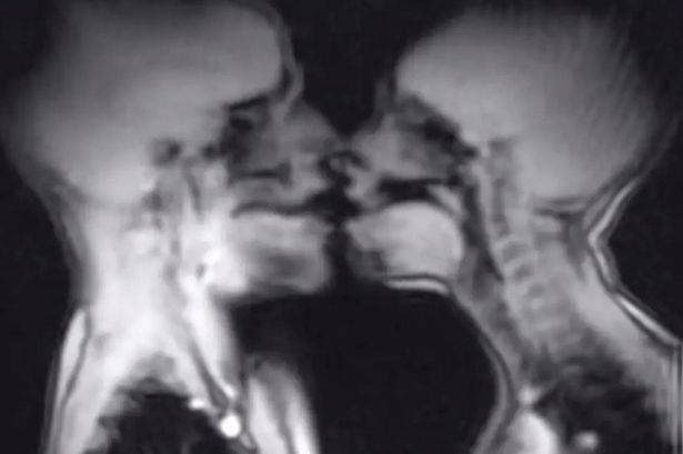
A unique scientific experiment has taken an unconventional approach to understanding human anatomy during sexual activity. In a study featured in the British Medical Journal from 1999, a couple, identified as Ida Sabelis and her boyfriend Jupp, participated in a magnetic resonance imaging (MRI) scan while engaging in sexual intercourse. The findings provided valuable insights, though one aspect of the results left researchers puzzled.
The study concluded that capturing magnetic resonance images of male and female genitals during coitus is not only feasible but also enhances the understanding of anatomical responses. Notably, the images revealed that during intercourse in the “missionary position,” the penis assumes a shape similar to that of a boomerang, with approximately one-third of its length comprising the root of the penis. Additionally, the researchers observed that during female sexual arousal, the uterus elevated and the anterior vaginal wall lengthened. Interestingly, the size of the uterus did not change during sexual arousal.
Despite these intriguing findings, a recurring phenomenon emerged during the experiment: the rapid filling of the female participants’ bladders. This observation, noted in all 13 instances of sex conducted in the MRI machine, baffled the researchers. Menko Victor ‘Pek’ van Andel, an expert involved in the study, proposed a hypothesis suggesting that this response could be an evolutionary mechanism to encourage women to urinate after sex, potentially reducing the risk of urinary tract infections. This theory, while not confirmed, was supported by the fact that each final scan consistently displayed a full bladder, even though many women had used the restroom prior to their MRI session.
Ida Sabelis, reflecting on her experience, stated that the MRI session was not particularly romantic but emphasized that their motivations were rooted in a desire to advance the understanding of female anatomy in scientific research. She described the experiment as an “act of love and a performance,” highlighting the significance of breaking barriers in women’s rights and health.
While the study provided important insights, it also serves as a cautionary tale regarding safety protocols during medical procedures. In a separate case, a 22-year-old woman suffered serious injuries after accidentally bringing a metal sex toy into an MRI machine. Believing it to be made entirely of silicone, she inserted a “butt plug” prior to the scan. The metal core of the toy reacted dangerously with the MRI’s magnetic field, resulting in the device being forcibly removed from her body. Reports indicate that the toy was pulled through her body into her chest cavity, leading to significant injuries, though she ultimately survived.
This incident prompted warnings about the importance of avoiding metal objects during MRI scans. A social media user, known as DreadPirateZero, shared a photo of the aftermath, advising others to refrain from using such items during medical appointments.
As researchers continue to explore the complexities of human anatomy and sexual health, studies like these underline the need for safety awareness and further investigation into the physiological responses observed during sexual activity.